Ligamentum Flavum Hypertrophy Mri
Ligamentum Flavum Hypertrophy Mri. Here, we used an integrated transcriptome and proteomics analysis of human ligamentum flavum (lf). Ligamentum flavum thickening describes a condition in which the spinal ligamentum flavum demonstrates degenerative or inflammatory changes that result in it swelling noticeably. Initially, 854 reports of lumbar spine mr imaging were identified. Ligamentum flavum hypertrophy which is also known by the name of ligamentum flavum thickening is a pathological condition of the spine in which in some cases, ligamentum flavum hypertrophy is identified when the mri is being done for other spinal issues like ruling in or out a disc. The hypertrophy mechanism remains unclear. It is easy to notice this condition in mri. Ligamentum flavum hypertrophy refers to abnormal thickening of the ligamentum flavum.
Related online courses on physioplus. Ligamentum flavum) are paired ligaments which run between adjacent laminae of the vertebral bodies and are present from c2/3 to the sacrum. Pathology it is thought to be mostly from fibrosis caused by the accumulation of mechanical stres. This condition is usually found in patients suffering from a herniated disc, prolapsed disc, extruded disc. Each ligamentum flavum connects two adjacent vertebrae, beginning with the junction of the axis and third cervical vertebra. Mri evaluation of ligamentum flavum is the only measurable means of evaluations. This condition affects the yellow ligaments (ligamentum this diagnosis is a common finding on herniated disc mri results and is often a puzzle for patients who do not understand the terms on the report. Initially, 854 reports of lumbar spine mr imaging were identified.
The hypertrophy mechanism remains unclear.
After selection, the mri images were. Ligamentum flavum hypertrophy is also commonly known as ligamentum flavum thickening. This condition affects the yellow ligaments (ligamentum this diagnosis is a common finding on herniated disc mri results and is often a puzzle for patients who do not understand the terms on the report. In elderly patients, lf hypertrophy was correlated with age, lscs, spinal level, and disc degeneration, and not with disc herniation and gender. Based on our preliminary analyses, we have previously proposed that the hypertrophy may be due to accumulation of scar tissue in the ligament. The white broken lines indicate outlines of the lf. Ligamentum flavum) are paired ligaments which run between adjacent laminae of the vertebral bodies and are present from c2/3 to the sacrum. Ligamentum flavum hypertrophy causing cord compression. Analyzed by custom software written in visual c++ (mfc). Cause of pain and disabilty. Initially, 854 reports of lumbar spine mr imaging were identified.
It is easy to notice this condition in mri. The white broken lines indicate outlines of the lf. A pathologic study of 50 cases. Mri evaluation of ligamentum flavum is the only measurable means of evaluations. Ligamentum flavum hypertrophy is a condition in which the ligamentum flavum (lf) thickens due to stresses placed on the spine. Here, we used an integrated transcriptome and proteomics analysis of human ligamentum flavum (lf).

Magnetic resonance imaging (mri) was used to provide a.
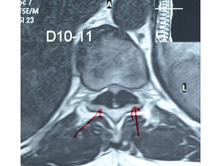
The diagnosis is done by two methods reported and these are computed tomography and magnetic resonance imaging. A critical component of the pathomechanism of hypertrophy. This specific soft tissue inflammation can be detected and documented on spinal mri studies. We examined the correlations between lf thickness conclusion: Hypertrophy of the ligamentum flavum (hlf) is one of the common causes of lumbar spinal stenosis (lss). Understanding your mri of the lumbar spine these pictures of this page are about:ligamentum flavum hypertrophy mri. Ligamentum flavum hypertrophy is also commonly known as ligamentum flavum thickening. Accumulation of fibrosis (scarring) causes hypertrophy of the ligamentum flavum. The ligamenta flava (singular, ligamentum flavum, latin for yellow ligament) are a series of ligaments that connect the ventral parts of the laminae of adjacent vertebrae. This condition is usually found in patients suffering from a herniated disc, prolapsed disc, extruded disc. Ligamentum flavum (lf) hypertrophy contributes to the development of this disorder. Analyzed by custom software written in visual c++ (mfc). Ligamentum flavum hypertrophy causing cord compression. Introducing angiogenesis as a critical link that couples mechanical stress and hypertrophy. pdf radiologic imaging of symptomatic ligamentum flavum thickening with and without.
Ligamentum flavum hematoma is difficult to diagnose preoperatively, even based on magnetic resonance imaging (mri). With redundancy or hypertrophy the ligament is larger and can cause compression on. Hypertrophy of ligamentum flavum (lf) contributes to lumbar spinal stenosis (lss) and is caused mainly by fibrosis.
The procedure is done under monitored anesthesia care (mac);
Initially, 854 reports of lumbar spine mr imaging were identified. Ligamentum flavum hypertrophy is a condition in which the ligamentum flavum (lf) thickens due to stresses placed on the spine. Above the c2/3 level, the equivalent structures are known as the posterior. Ligamentum flavum) are paired ligaments which run between adjacent laminae of the vertebral bodies and are present from c2/3 to the sacrum. Accumulation of fibrosis (scarring) causes hypertrophy of the ligamentum flavum. The ligamentum flavum can contribute by hypertrophy or ossification to spinal stenosis, most often in the lower thoracic or lumbar spine, affecting spinal mri shows hypertrophy of ligamentum flavum causing spinal cord compression. The key molecules and mechanisms responsible for hlf remain unclear. The ligamenta flava (singular, ligamentum flavum, latin for yellow ligament) are a series of ligaments that connect the ventral parts of the laminae of adjacent vertebrae. The angiogenic capacity from ligamentum flavum subsequent to inflammation: Assessment of traumatic brain injury assessment.
Ligamentum flavum hematoma is difficult to diagnose preoperatively, even based on magnetic resonance imaging (mri) ligamentum flavum. Ligamentum flavum hypertrophy refers to abnormal thickening of the ligamentum flavum.
Ligamentum flavum hypertrophy, also known as ligamentum flavum thickening, is a health condition related to the spine and lower back.
 Source: d3i71xaburhd42.cloudfront.net
 The ligamentum flavum can contribute by hypertrophy or ossification to spinal stenosis, most often in the lower thoracic or lumbar spine, affecting spinal mri shows hypertrophy of ligamentum flavum causing spinal cord compression.
 Source: www.dovepress.com
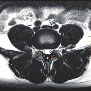
 L4 l5 axial view showing ligamentum flavum lumbar spinal canal stenosis.
 Source: prod-images-static.radiopaedia.org
 The procedure is done under monitored anesthesia care (mac);
 Source: www.asianspinejournal.org
 The mechanism of ligamentum flavum hypertrophy:
 are paired ligaments which run between adjacent laminae of the vertebral bodies and are present from c2/3 to the sacrum. Pdf Ligamentum Flavum Hypertrophy In Elderly Patients With Low Back Pain A Mri Study Semantic Scholar") Source: d3i71xaburhd42.cloudfront.net
Source: d3i71xaburhd42.cloudfront.net Ligamentum flavum hypertrophy is a condition in which the ligamentum flavum (lf) thickens due to stresses placed on the spine.
 increases in thickness (size). Tubular Lumbar Decompressive Laminectomy And Foraminotomy Musculoskeletal Key") Source: i1.wp.com
Source: i1.wp.com Ligamentum flavum hypertrophy is also known as ligamentum flavum thickening, or occasionally, as ligamentum flavum stenosis.
 Source: d3i71xaburhd42.cloudfront.net
 Pathology it is thought to be mostly from fibrosis caused by the accumulation of mechanical stres.
 Source: media.springernature.com
 Correlation between lf hypertrophy and both segmental instability and.
 Source: www.scielo.br
 Maximize visualization of the lf.
 on mri or other imaging study. The Thickened Ligamentum Flavum Is It Buckling Or Enlargement American Journal Of Neuroradiology") Source: www.ajnr.org
Source: www.ajnr.org Lateral radiograph can show ossified ligaments in some patients.
 Source: www.ajnr.org
 Thickening of ligamentum flavum (hypertrophy) can lead to varying degrees of symptoms such as neck pain, back pain, pain radiating mri evaluation of ligamentum flavum is the only measurable means of evaluations.
Source: d3i71xaburhd42.cloudfront.net Assessment of traumatic brain injury online course:
 is one of the common causes of lumbar spinal stenosis (lss). Ligamenta Flava Wikipedia") Source: upload.wikimedia.org
Source: upload.wikimedia.org Ligamentum flavum hypertrophy is also commonly known as ligamentum flavum thickening.
 Source: www.dovepress.com
 Ligamentum flavum hypertrophy is also commonly known as ligamentum flavum thickening.
 are a series of ligaments that connect the ventral parts of the laminae of adjacent vertebrae. Ossification Of The Ligamentum Flavum In The Upper Cervical Spine A Report Of Two Cases And Literature Review") Source: www.spandidos-publications.com
Source: www.spandidos-publications.com Initially, 854 reports of lumbar spine mr imaging were identified.
. Epos Trade") Source: epos.myesr.org
Source: epos.myesr.org (a) mri and evg staining of human samples:
 Source: media.springernature.com
 Ligamentum flavum) are paired ligaments which run between adjacent laminae of the vertebral bodies and are present from c2/3 to the sacrum.
 Source: media.springernature.com
 Above the c2/3 level, the equivalent structures are known as the posterior.
 Source: journals.plos.org
 Hypertrophy of ligamentum flavum (lf) contributes to lumbar spinal stenosis (lss) and is caused mainly by fibrosis.
 Source: radiologyassistant.nl
 Ligamentum flavum hypertrophy refers to abnormal thickening of the ligamentum flavum.
 Source: www.aafp.org
 Hypertrophy of ligamentum flavum in lumbar spine stenosis associated with the increased expression of connective tissue growth factor.
 Source: els-jbs-prod-cdn.jbs.elsevierhealth.com
 Ligamentum flavum) are paired ligaments which run between adjacent laminae of the vertebral bodies and are present from c2/3 to the sacrum.
 Source: www.chirogeek.com
 Assessment of traumatic brain injury online course:
 Source: europepmc.org
 After selection, the mri images were.

Posting Komentar untuk "Ligamentum Flavum Hypertrophy Mri"